Juniper Publishers - Flexible Intramedullary Titanium Elastic Nailing of Fracture Shaft of Radius and Ulna in Children at a Tertiary Care Teaching Hospital
Orthopedics and Rheumatology Open Access Journal
Abstract
Introduction: Displaced fracture shaft of both bone forearms in children can still managed with close reduction and cast application. If, it has failed or remain inadequately reduced after closed reduction require intramedullary fixation to achieve functional outcome. This study assesses the functional outcome of treating displaced fracture shaft of both bone forearm in children with intramedullary flexible titanium elastic nailing.
Method: 79 children aged 3 to 15 years with displaced fracture of shaft of both bone forearm underwent flexible titanium elastic nail. The patients were followed up for a period of 12 months.
Results: Close reduction followed by nailing was possible in 71 patients, while 8 patients required open reduction through mini incision of both the radius and ulna fracture prior to nailing. 74 patients had excellent results and 5 patients had good results. 13 patients had minor complications including skin irritations over prominent hardware, superficial nail insertion site infection were noted in our study. 2 patients had a restriction of 20° of pronation and 10° of supination, 2 patients restriction of 15° of pronation and 1 patient had 8° volar angulation at the radial bone with limitation of 5° supination. All fractures were united in acceptable alignment by an average 9 weeks and nails were removed at an average of 6 months.
Conclusion: Flexible nailing leads to more versatile and efficient application of internal fixation for fracture shaft of both bone forearm, which permits early mobilization and return to the normal activities of the patients, with very low complication rate.
Keywords: Shaft; Radius and ulna; Forearm fractures; Intramedullary; Flexible titanium nailing.
Abbreviations: FIN: Flexible Intramedullary Nailing; AP: AnteroPosterior; IERC: Institutional Ethical Review Committee; ORIF: Open Reduction and Internal Fixation; ESIN: Elastic Stable Intramedullary Nailing; TEN: Titanium Elastic Nails; SPSS: Statistical Package for Social Sciences.
Introduction
Being so complex and important in relation to function of the upper extremity, injuries of the forearm can result in potentially hazardous consequences. There is no doubt that forearm shaft fractures are potentially harmful and challenging to manage [1]. Displaced fractures of the forearm shaft in children can be reduced and can still be managed with cast, but there is a relatively high incidence of redisplacement, malunion and consequent limitation of function. Union is rarely a problem [2,3]. The most common indications for surgery are failure of closed reduction, open fractures, and fracture instability. In these situations, if left untreated, malunion is more likely to occur, which will disturb the function of the upper extremities [4]. Shoemaker et al. suggested that the ideal mode of fixation of pediatric forearm fractures should maintain alignment, be minimally invasive and inexpensive and carry an acceptable risk profile [5]. As compared to intramedullary fixation, ORIF with plates and screws has got several disadvantages such as large incisions with poor cosmesis, more soft tissue dissection, higher incidence of infections and difficult removal. As far as Intramedullary fixation is concerned there are several implants such as k-wires, Steinmann pin and rush nails but they have their own disadvantages such as Kirschner wires and Rush nails are rigid and difficult to insert through the metaphysis of children’s bones. Because these disadvantages, flexible intramedullary nail (TENS) were devised to overcome this problem which produces a three-point fixation to maintain bony alignment and now become very popular method for managing forearm fractures in children [6].
The flexibility of flexible intramedullary nail (TENS) allows for micromotion at the fracture site is caused by the elasticity of the nail, which are thought to be beneficial for rapid fracture healing [7,8]. This elastic deformation within the medullary canal creates a bending moment within the long bone that is not rigid, but that is stable enough to reduce and fix the fracture [9]. Hence, the current study is intended to review retrospectively the clinical, radiological and functional outcome of Flexible Intramedullary Titanium Elastic Nailing of Fracture Shaft of Radius and Ulna in Children at a Tertiary Care Teaching Hospital.
Methods
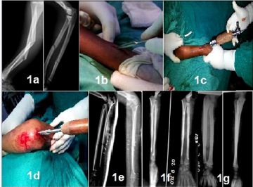
In this study, the data covered 741 consecutive paediatric forearm both-bone shaft fracture cases were attended at Department of Orthopaedics, National Medical College from 2013 to 2015. Among 741 children, we report the outcome of flexible intramedullary nailing (FIN) for diaphyseal fractures of the forearm in 79 children aged between 3-15 years during a 3 year period with inclusion criteria of displaced forearm shaft fracture(angulated >20° and displaced >50%) (Figure 1a) or grossly rotated fractures, re fractures, failed close manipulation, open fracture (Gustilo and Anderson class I and II). Patients with isolated forearm bone fracture, floating elbow, pathological fracture, open fracture GA type III or fractures with neurovascular injury were excluded. Shaft fractures with associated disruption of the radiocapitellar joint (Monteggia fracture and equivalents) and the distal radioulnar joint (Galeazzi fracture and equivalents) were excluded. The diagnosis was made on x-ray involving both radius and ulna. Fractures were temporarily stabilized with back slab in the emergency or outpatient department; patients were admitted in ward and started on regular analgesia. The patient’s family was counseled for the surgical procedure and informed consent was taken.
Demographics data of patient including age, sex, mechanism of injury, site of fracture and type of fracture, fixation method and indications for surgery were documented (Table 1 & 2). Complication rates, time for fracture union and final range of motion were evaluated in subsequent follow up. Radiographic union was defined as bony trabeculae traversing the fracture on anteroposterior (AP) and lateral radiographs [8]. In (Table 1), final outcome was measured on 12 months on the basis of grading system by Price et al. [10]. Good to excellent outcome were taken as acceptable outcome. The results were considered excellent if no complaints with strenuous physical activity or a loss of pronation / supination of < 10° good if mild complaints with strenuous activity and/or 11°-30° loss of forearm rotation; fair if subjective complaints during daily activities or 31°-90° loss of forearm rotation and all other results were considered poor [5]. Data were analyzed using SPSS/PC+V. 6.0 (Statistical Package for Social Sciences, SPSS Inc version 15, Chicago, Illinois) [11]. The results were shown in frequencies and percentages. Functional outcome was compared among age groups, gender and timing of presentation. Chi square test was used and p value of < 0.05 was taken significant. All were part of an ongoing study approved by the Institutional Ethical Review Committee (IERC) for research on human subjects at National Medical College, Nepal.
Surgical technique
Patient was placed supine on the operation table under general anesthesia. Tourniquet was applied in the arm with proper padding in case an open reduction is needed. Image intensifier was adjusted to obtain appropriate AP and lateral views of forearm. A flexible nail size 2.0 to 3.0 mm was used for either bone. Proper size flexible nail as decided preoperatively by measuring the medullary canal in X-ray was used. The diameter of bone in anteroposterior added with diameter in lateral radiograph divided by 2 approximately gave the size of nail to be used. We used nail of 0.5 mm smaller than the calculated size. The radial and ulna nail with its proximal 5mm pre-bent was bent to about 15 to 30 degrees for easy passage of the pin through the medullary cavity. Because the radius is often more difficult to reduce, it was splinted first.
The radial bone is approached through one cm longitudinal incision performed on the lateral side of the distal metaphysis carefully avoiding damage to the superficial branch of the radial nerve. A hole is drilled in the bone with an awl, first perpendicularly and then obliquely towards the elbow just proximal to the distal radial epiphysis (Figure 1b). Optimal care was taken not to injure extensor tendons and superficial radial cutaneous nerve. Then an appropriate size flexible intramedullary nail with its proximal 5mm pre-bent at 30° is introduced with the inserter for TEN into the medullary canal, with the nail tip at right angles to the bone shaft and pushed retrograde with a hammer if necessary, to the fracture site (Figure 1c). Then rotate the nail through 180° with the inserter and align the nail tip with the axis of the medullary canal.
Figure 1: grossly rotated fractures.
The fracture was reduced by external manipulation and the pre bent nail was pushed proximally and advanced through fracture site with oscillating movements and was stopped short of the physis, at the level of bicipital tuberosity. The curved tip of the nail can be used as an aid to reduction of the fracture. If an acceptable reduction cannot be obtained, then open reduction through limited approach and intramedullary fixation is performed. The distal end of the nail was bent and cut 5-10 mm from the bone. The skin was closed with one or two stitch. For the ulna fracture, the stab incision was made on the proximal end of forearm over lateral surface of the olecranon 2cm distal to physis under fluoroscopy. An awl was introduced to make an oblique entry hole in the ulna 2cm distal to olecranon physis (Figure 1d). The point of entry was easily palpable and proximal to the annular ligament and head of the radius. The nail which was pre bent at 30° at the tip was antegradely introduced with the inserter for TEN in the oblique entry hole and gently negotiated and pushed distally. Fluoroscopy was used during reduction. If required, fracture site was exposed by a small incision and reduction accomplished. The bent tip of nail aided in the reduction and the nail was pushed into the distal ulna, stopping short of the physis under the guidance of fluoroscopy. The nail was cut close to the bone, leaving enough ends for easy removal later but without any tenting the skin. The skin was closed over the cut end. Skin over the incision made for exposing the fracture, if given, was closed too.
Stability of the fracture was accessed postoperatively (Figure 1e). If the fracture was stable then simple sling is applied for 5 days, if fracture was unstable then long arm posterior plaster slab was applied for at least 4 weeks. The patient was instructed to avoid excessive loading of the involved limb until adequate callus formation is observed on radiographs made at approximately four weeks and is advised to refrain from sports for 6-8 weeks. Physiotherapy was started as early as possible. Active and passive range of motion exercises for fingers were started on 1st post operative day. Intermittent extension and flexion of elbow and wrist was allowed from second post operative day as tolerated by the patient if the patient was in sling.
Patients were discharged in 2nd day on continue of nonsteroidal analgesic medication and oral antibiotics for 3 days when clinically stable and pain is minimal. Stitches were removed on 10th post operative day in outpatient department and splint was continued depending on stability of fracture for more comminuted fractures. It was continued up to 3 weeks otherwise it was discarded after 10 days and active movements were encouraged. Supination and pronation was allowed only after six weeks. Patients were followed up at 15 days, three weeks, four weeks, six weeks, three months, six months, nine months and 12 months time for clinical radiological evaluation of union and functional outcome. Absence of tenderness and mobility at fracture site is an indication for normal activities.
Results
An outcome of flexible intramedullary nail fixation of 79 patients with shaft fractures of both bone forearm in children is undertaken in the study. Mean age was 10.4 ± 3.09 years (range 3 to 15 years). There were 52 male patients (65.8%) and 27 females (34.2%). Right forearm was fractured in 46 children (58%) and left forearm was fractured in 33 children (41%). Out of 79 forearm fractures, 65 were closed fractures (82%) and 9 were open Gustillo Anderson type I fractures (12%) and 5 were open Gustillo and Anderson type II fractures (6%). Transverse fractures accounted for 46(58%) cases, communited fractures 11(14%), oblique fractures 16(20%) and spiral fractures 6(8%). Among 55(69.6%) of middle third forearm shaft fracture, 44(55.6%) of the patients were males and 11(14%) females (P< 0.001). Among these 55 patients 41 were among age group of 3-10 years and 14 were among age group of 11-15 years.
Among fourteen patients (17.7%), eight (N=8) proximal third forearm shaft fractures were seen in male 10% and 8% females (N=6). Among these 14 patients, four were among age group of 3-10 years and ten were among age group of 11-15 years. Ten of the patients (12.6%) with distal third forearm shaft fracture were male 8(10%) and 2 females (2.5%). All these patients were among age group of 3-10 years. Shortening was seen in 38% of the middle-third shaft of forearm fracture cases (N=21). The typical presentation is demonstrated on the radiograph in (Figure 1a).
Most (70%, N=55/79) of the middle-third shaft of forearm fractures showed angular deformity primarily in radiographs at admission. Angular deformity of >15º was seen in half of 55 (51%, N= 28/55) middle third shaft of forearm fractures, one fifth (20%, N=11/55) showed 0-15 degrees of angular deformity and a third (29%, N=16/55) had no deformity primarily in the study. In cohort, over 2 mm of displacement was seen in 32% (N=25/79) of the patients with a forearm shaft fracture. Two thirds (68%, N=54/79) showed < 2 mm of displacement primarily. Among 55 children of both bone forearm shaft fracture, initial angulation of the radius fracture in 8 children ranged from 10° to 50° in the lateral plane and 10° to 32° in the anteroposterior plane (Figure 1a). The Initial angulation of the ulna fracture in 4 children ranged from 12° to 67° in the lateral plane and 5° to 36° in the anteroposterior plane.
The (Table 3) show, the initial angulation of both the radius and ulna fracture in 43 children ranged from 10° to 67° in the lateral plane and 10° to 32° in the anteroposterior plane. There were no significant differences in angulation between the fractures of both bones and angulation of radius and ulna fracture (p>0.5). 46 children sustained injury by a fall on outstretched hand while playing (58%) and 6 children suffered injury in a road traffic accident (7%). One fifth (19.3%, N= 15) of all shaft fractures were sports related. Other mechanisms of injuries included fall from height (15%, N=12). 67(84.8%) children out of 79 were brought to the hospital within 24 hours of injury. A few (N=12, 15%) were admitted one day later. Most of the patients presented within 3 days of injury. Mean duration of injury was 3.39±1.38 days with range from 2 to 6 days.
All the patients were prepared and operated as early as possible once the general condition was stable and the patient was fit for surgery. Among 79 children, closed reduction and internal fixation of fracture shaft of both bone forearm under guidance of image intensifier with flexible intramedullary nail was accomplished in 71(90%) patients. However, 8(10%) patients required limited open reduction and internal fixation (ORIF) with flexible nail because of difficult cannulation due to soft tissue interposition. The average duration between trauma and surgery was 3.96 days and average duration of surgery is 59.9 minutes. The duration of surgery (from incision to closure of wound) was less than or equal to 1hours in majority of cases 73(92%) and all these patients had closed reduction and internal fixation.
The operating time was more than 1hour in 6(8%) patients and all these patients had open reduction and internal fixation. The mean hospital stay was 2.5 days (1 to 5) 85% were discharged within three days. Postoperative, 90% of the patients were applied simple sling for 5 days; where as in a few 10% of patients were immobilized with long arm posterior plaster slab for 4 weeks. In early postoperative period, no patient had compartment syndrome or nerve palsy. Patients were followed up for mean duration of 11months (range 6-12 months). Union was achieved in all the patients. No cases of nonunions or malunions were reported.
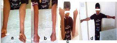
There was evidence of radiological union of fracture at 6 weeks in 8(10%) patients, 9 weeks in 63(80%) patients and 12 weeks in remaining 8(10%) patients (Figure 1f). The average time for union of fracture was 9 weeks. At final follow up of 12 months, excellent acceptable functional outcome according to Price et al. [10] criteria was found in most of the patients (n=74 (94%) followed by good in 5(6%) patients and there were no poor outcome cases (Table 4). Among patients with good outcome, two patients had a restriction of 20° of pronation and 10° of supination, two patients restriction of 15° of pronation and a 14-year-old boy with 8° volar angulation at the radial bone and limitation in supination of about 5° upon final follow-up. The remaining patients had a range of movement of the elbow, forearm and wrist within 5° of that of the normal side (Figure 2a-2d). At final follow up, no patient had any pain at fracture site even with strenuous activities like participation in sporting activities. Out of the 79 patients, thirteen (16%) patients had minor complications; six patients had nail insertion site skin irritation over prominent ulnar hardware, backing out of ulnar nail requiring early removal in one, two had superficial nail insertion site infection which was successfully treated with oral antibiotics and daily dressing for ten days and superficial skin break down with exposed hardware requiring nail removal under local anaesthesia in four cases. The nail removal was performed under general anesthesia at an average of six months (range 4-9 months) (Figure 1g)). The mean interval between the initial surgery and removal of the nail was 3.2 months. The average operation time for removal of the nail was 22 minutes. There was no incidence of re fracture after removal of nail.
Figure 2: Remaining patients having range of movement of the elbow, forearm and wrist within 5° of that of the normal side.
Discussion
Up to 25% of complete forearm fractures displaced during the follow-up and may require a second intervention [12]. Several authors have suggested that a reduction is unacceptable if the patient has an angular deformity >10° or complete displacement [5,13]. Parameters for accepting rotational malalignment range from 30°-45° to none and some authors have noted that rotational remodeling is not predictable [5,14-16]. The remodeling capacity depends on age, the site of fracture, the direction of angulation and its magnitude. Rotational deformity does not remodel [2]. The most challenging forearm fractures are both-bone fractures in the middle-third of the shaft [17] and those that are proximal do not remodel as predictably; therefore, these require a more anatomic reduction [18]. It has been reported that middle third fractures cause more functional limitations compared to distal third diaphyseal forearm fractures [19,20]. A cadaver study showed that supination losses were much more obvious than pronation losses in middle third forearm fractures [20].
In our study most 70% (55/79) of the middle third shaft of forearm fractures treated with flexible intramedullary nail in which showed angular deformity >15º in radiograph at the time of admission. Daruwalla et al. [4] recommended operative intervention for midshaft and proximal forearm fractures with angulations >10° because of limited remodeling potential in these areas of the bone. Matthews LS et al. [21] showed in a cadaveric study that forearm angular deformities of 10° will not result in significant loss of forearm pro/supination but that angulation of 20° will restrict forearm rotation approximately 30%. Another cadaveric study by Tarr RR et al. [20] demonstrated that fracture angulation between five and ten degrees at mid shaft of forearm can lead to pronation deficit of 5% to 27% of normal. Given the potential failure of non operative management (1.5% to 31%) and the importance of minimizing angular deformity to preserve normal forearm rotation, operative management of pediatric forearm fracture has been increasingly popular.
Flexible intramedullary nailing is preferred fixation method for pediatric forearm fractures with few reported complications [22-24]. Most series show good to excellent results using this method [1,5,13,15,21]. Flexible intramedullary nails as shown in the present study where 74(94%) patients had excellent results and is comparable with other similar study (Table 4). In comparision to platings which is more suitable for older children (10-15 years) for accurate anatomic alignment and early unprotected range of motion, intramedullary flexible nail also offer various potential benefits in terms of cosmesis, easy removal of implants after treatment and decreased chances of neurovascualr injuries [16,25]. Intramedullary fixation promotes rapid union, reduces the risk of infection, synostosis and avoids unsightly incisions that are necessary for plate fixation and hardware removal [1].
Intramedullary flexible nail (Titanium elastic nails) comes in a range of sizes with both variable length and diameter allowing optimal implant choice for each patient. Kirschner wires and Rush nails are rigid and difficult to insert through the metaphysis of children’s bones. Flexible intramedullary nail (TENS) were devised to overcome this problem which produces a three-point fixation to maintain bony alignment, first at their entry point of metaphysis; second the contact of the apex of the nail with the inner wall of the cortex at the opposite side of the intramedullary canal preferably close at the site of the fracture; and third the anchoring of the tip of the nail into the opposite metaphysis of the other end of the bone [6]. The flexibility of flexible intramedullary nail (TENS) allows for micromotion at the fracture site, which are thought to be beneficial for rapid fracture healing and enhance callus formation [7,8]. This micromotion is caused by the elasticity of the nail. This elastic deformation within the medullary canal creates a bending moment within the long bone that is not rigid, but that is stable enough to reduce and fix the fracture [9]. The curvature of the nail is achieved by pre-bending them beyond their elastic limit. End-to-end reduction helps control rotational alignment and limited motion at the fracture site promotes the formation of external callus by converting shear stress at the fracture site into fracture compression [26].
Flynn JM et al. [26] reported a single bone fixation is technically easier and involves less operating time; stabilisation of ulna prevents the development of a cosmetically unacceptable bow and provides a fulcrum against which the radius can be maintained in an improved position. However, redisplacement of the non fixed bone and loss of reduction may occur [5,23]. Kapoor et al. [8] observed secondary displacement in three cases out of five, where a single bone was nailed in a both bones forearm fracture, one such case resulting in a symptomatic distal radioulnar joint subluxation due to 12° of ulnar angulation [8]. Lascombes et al. [26] recommend intramedullary nail fixation of both radius and ulna. Forearm bones are bound together by the annular ligament above, the triangular ligament below and the interosseous membrane in between. Therefore, the osteosynthesis has to include both bones since the nailing of a single bone may lead to the displacement of the second bone.
In our study included flexible intramedullary nail fixation of fracture shaft of both radius and ulna to prevent secondary displacement. In both bone forearm fractures we now routinely stabilise both the bones even if one of the fractures is undisplaced. Amit et al. [22] described the results of treatment of 20 unstable diaphyseal fractures of the forearm in adolescent patients treated with closed intramedullary nailing and favored that technique rather than plate fixation because of the appropriate reduction, reduced complication rate, negligible cosmetic defect and the ability to perform rod removal under local anesthesia. Stanley and Wilkins et al. [18] reported on 50 patients with mid shaft fractures of the radius and ulna treated with closed reduction and percutaneous intramedullary pinning. Reduction was achieved through a limited open approach to one or both bones in their first six patients.
Pogorelic Z et al. [27] reported in 37 patients the forearm both bone fracture was reduced by closed means, whilst in the other 15 open reduction was required due to difficulty in reduction and soft tissue interposition. In our study, 71(90%) patients were treated with closed reduction and internal fixation of fracture shaft of both bone forearm under guidance of image intensifier with flexible intramedullary nail and only 8(10%) patients needed mini open reduction due to soft tissue interposition to pass the nail across the fracture site. This is comparable to studies by Richter et al. [15] (closed reduction 84%) and Alam W et al. [28] (closed reduction 72%). Cullen et al. [23] (open reduction 75%) and Luhmann et al. [29] (open reduction 50%). Close reduction or open reduction before intramedullary nailing yield similar functional results, with similar complication profile in pediatric diaphyseal fracture [30, 31]. Though we did not compare the results of open Vs close technique but we included both techniques, where results are good to excellent.
In our study average time for union of fracture was 9 weeks whereas the average time for union of fracture was 7 weeks in a study conducted by Kapoor V et al. [10] on forearm fractures in children treated by elastic stable intramedullary nail. The difference could be due to our follow up interval which was at three weeks, four weeks, six weeks, nine weeks, three months, six months, nine months and 12 months. At six months, no patient had any pain at fracture site even with strenuous activities like participation in sporting activities. These findings are in accordance with study by Ali AM et al. [32]. At six months none had loss of movement at wrist and elbow. This is in accordance to study conducted by Fernandez F et al. [25] which was done to compare outcome between plating and nailing (ESIN) in forearm fractures in children where in there was no restriction of movement at wrist and elbow in group of patients managed by ESIN.
In this study nail removal was performed at an average of six months. There was no incidence of re fracture after removal of nail. This is in accordance to study conducted by Slongo et al. [33] who recommended that nails should not be removed before 4-6 months and the fracture must be consolidated to minimize the risk of refracture. Manish C et al. [34] reported, refracture will occur even in the presence of in situ nails and therefore there seems to be little point in leaving the nails any longer that the time required for union and consolidation.
Shivanna et al. [35] reported 45 children aged 5-15 years with displaced diaphyseal forearm fractures treated titanium elastic nailing and immobilized postoperatively with an above-elbow plaster slab for 2 weeks till the swelling is completely resolved followed by encouraging range of motion exercises. Due to recurring instability, many clinicians recommend routine post operative casting in forearm fracture in children treated by flexible intramedullary nail [1,15]. Hence, in our study 90% of the patients were placed in a simple sling for 5 days and were allowed to early mobilization of the upper extremity as tolerated. This allowed in our patients with reduction in hospital stay, rapid return to daily activities, an ability to participate fully in school activities and avoidance of the discomfort and inconvenience of cast immobilization. Only 10% of patents were found unstable which was immobilized long arm posterior plaster slab for 4 weeks which is comparable to Alam W et al. [28] immobilized their patients for a period of 4 weeks. Luhmann et al. [29] who in their study immobilized children for a mean period of 7 weeks.
The procedure of inserting intramedullary nails is not without the possibility of complication. In this series of patients, we have reported a complication rate of 16%. This is similar to the complication rate reported by Lascombes et al. [36] and Parajuli NP et al. [37]. Yalcinkaya M et al. [30] reported complications rate ranged from 4-38% in patients treated with intramedullary nailing and Flynn JM et al. [13] showed that the overall complication rate in patients undergoing intramedullary nailing was 14.6%. The most common complication occurring in their series were delayed union, compartment syndrome, infection, skin irritation by hard ware and pin back out.
Berger P et al. [31] reported superficial infection at entry point of nail in 6% (2 out of 30) patients with no report of deep infection, compartment syndrome, nerve palsy, nonunion, malunion, refracture or nail migration. (Table 4) is a detailed analysis of functional outcome of the patient was done on the basis of following criteria by Price et al. [10]. In our study average age was 10.04 years and excellent outcome was observed in 74(94%) and good in 5(6%). No Poor results were observed in our study. Among thirteen (16%) patients of minor complication, reported six patients had nail insertion site skin irritation, backing out of ulnar nail requiring early removal in one, two had superficial nail insertion site infection and superficial skin break down with exposed hardware in four cases. No nonunions or malunions occurred. There were no deep infections noted. There was no reported loss of reduction after initial fracture fixation and no reported long-term complications with forearm rotation. Our results are comparable with that of Parajuli NP [37] who had excellent result was reported in 47(94%) patients while good in 3(6%) patients and eight (16%) patients were reported to have minor complications including skin irritation, back out of ulnar implant and skin breakdown with exposed implant.
Ahmad I et al. [38] reported the results of 22 patients with an average age of 9.5 years having unstable radius ulna forearm fractures. They observed excellent outcome in 18(82%) patients, good in 2(9%) and fair in 2 (9%) patients. Richter D et al. [15] who had 24(80%) patients with excellent results, 5(16.6%) with good results and 1(3.3%) with fair results with no poor results noted. In 1998 Luhmann SJ et al. [29] reported excellent results in 21(84%), good results in 4(16%) and no fair/poor results seen. Van der reis WL et al. [39] reported Excellent results in 18(78%) patients, Poor results in 5(22%) patients. Cullen MC et al. [23] reported Excellent results in 17(89.4%) patients, Good results in 2(10.5%) patients with no poor results noted. Lascombes P et al. [26] reported excellent results in 92% patients treated with elastic stable intramedullary nailing (ESIN), 8% good results with no poor results. Altay M et al. [40] reported Excellent results in 83.3%, good in 12.5%, fair in 2.1% and poor results in 2.1% patients. In the pediatric patient, non-union has not been reported in the literature, and good/excellent functional results are reported in nearly 95% of cases [22,23,26,41]. These excellent clinical results support the use of this technique in the operative treatment of displaced both bone forearm fractures in the pediatric patient.
Conclusion
Flexible nailing leads to more versatile and efficient application of internal fixation for fracture shaft of both bone forearm, which permits early mobilization and return to the normal activities of the patients, with very low complication rate, high union rates and excellent functional outcome. We, therefore recommend this surgical procedure for the treatment of displaced shaft of both bone forearm fractures in children.
To Know More
About Orthopedics and
Rheumatology Open Access Journal Please click on:
To Know More About Open
Access Journals Please click on:


Comments
Post a Comment